
A chemical clue carried by light
Silver can help scientists read a chemical clue carried by light. When laser light scatters from molecules, a small part changes energy as it interacts with their vibrations. The resulting pattern, called a Raman spectrum, can help reveal what is in a sample.
That pattern is sometimes described as a molecular “fingerprint.” It can be extremely faint. Molecules on or very near specially structured silver can produce much stronger signals, making small amounts easier to study. The method is called surface-enhanced Raman scattering, or SERS.
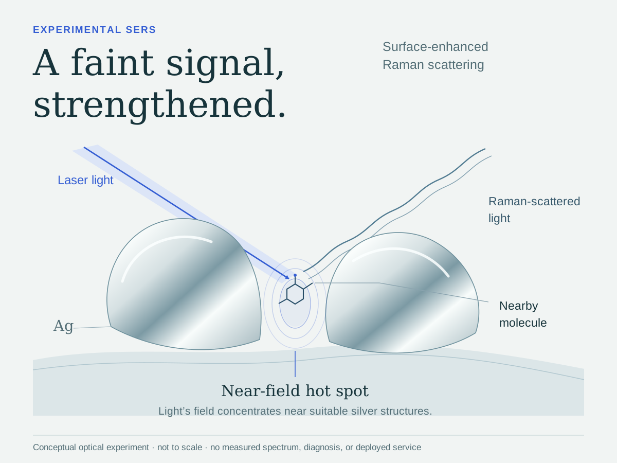
Small structures, a much stronger signal
Light can make electrons in the metal move together. These collective motions, called surface plasmons, concentrate the light’s electric field near the silver. Tiny regions with especially strong enhancement are known as hot spots.
Under suitable experimental conditions, silver surfaces can strengthen Raman signals by a million-fold or more. The size of the effect varies greatly with the surface, light and molecule. Even chemical changes at the silver surface can weaken it: a NIST study found that an oxide layer, made of silver and oxygen, sharply reduced the enhancement in its tests.
From a 1974 experiment to research today
In 1974, Southampton researchers recorded Raman spectra from pyridine, a chemical compound, on a roughened silver electrode. That experiment became an important part of SERS history.
Researchers now explore silver-based SERS for trace-chemical sensing and biological-sample analysis, including cancer-related studies of serum, the liquid remaining after blood clots. A stronger signal still needs careful interpretation and validation. These experiments do not establish routine hospital screening or airport deployment, and silver is not a universal detector or always a better choice than gold.
About this edition
Original source and editorial notes
Adapted from original material by Rich Silver. Original source card: Silver fact: supplied original card (publication date not established).
Reviewed 2026-11-05. This edition separates demonstrated SERS mechanisms and experimental sensing from routine clinical or airport use. Stronger signals depend on measurement conditions and do not guarantee identification. Reader-first revision explains Raman patterns and the fingerprint analogy before naming SERS or plasmons. Retains condition-dependent enhancement, the 1974 silver experiment, and the distinction between research results and validated identification/deployed screening. Does not introduce diagnostic performance, a universal detector claim or practical trace-detection instructions.
Sources & further reading
- Measurement of the distribution of site enhancements in surface-enhanced Raman scattering ↗
- SERS nose arrays based on a signal differentiation approach for TNT gas detection ↗
- Super-Resolution Imaging of Plasmonic Nanostructures ↗
- Effect of oxidation on surface-enhanced Raman scattering activity of silver nanoparticles: a quantitative correlation ↗
- Detection of explosives based on surface-enhanced Raman spectroscopy ↗
- Raman spectra of pyridine adsorbed at a silver electrode ↗
- Portable and Reliable Surface-Enhanced Raman Scattering Silicon Chip for Signal-On Detection of Trace Trinitrotoluene Explosive in Real Systems ↗
- SORS ↗
- Multi-cancer early detection based on serum surface-enhanced Raman spectroscopy with deep learning: a large-scale case–control study ↗