
Silver helped make a cell stand out
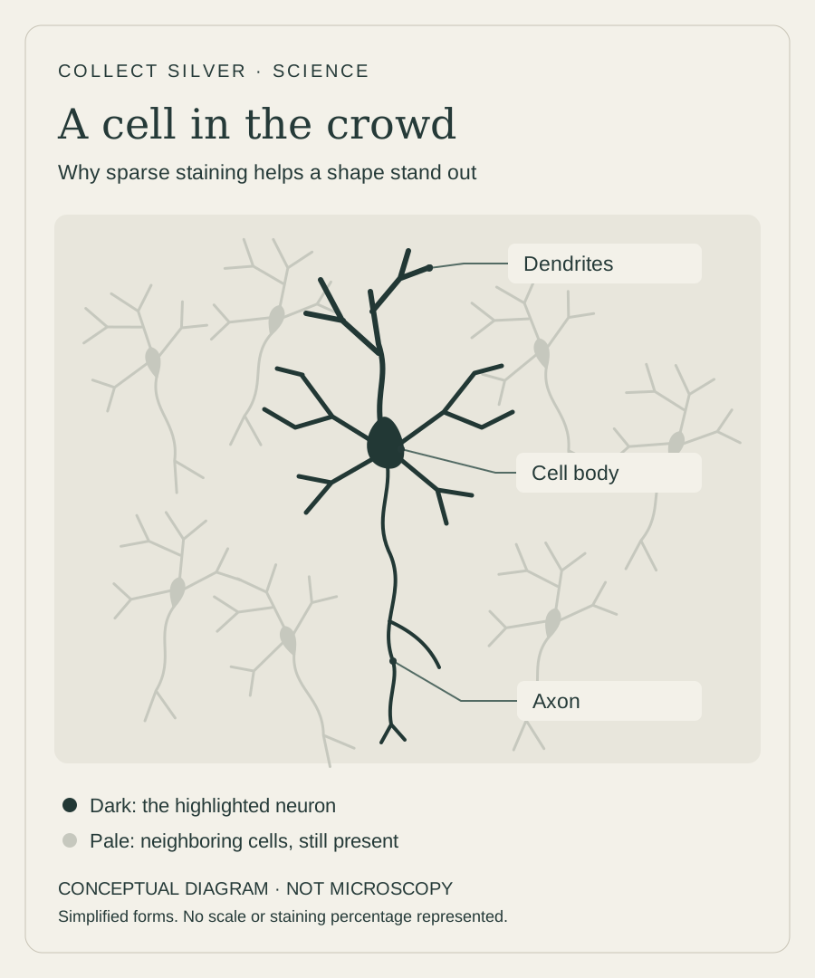
Silver helped scientists pick out individual nerve cells in a crowded microscopic scene. Picture a tree with all its branches tangled into the trees around it: tracing one branch through the thicket is a little like the problem nineteenth-century scientists faced when examining nervous tissue.
In 1873, Italian physician Camillo Golgi developed a staining technique that made individual nerve cells stand out in remarkable detail. Earlier methods had already revealed parts of neurons, but tracing their delicate extensions could be difficult. Golgi’s “black reaction” offered a much clearer view of their shapes.
The black in the black reaction
The crucial ingredient was silver nitrate. In tissue treated with potassium dichromate, it produced deposits of silver chromate, a silver-containing compound. The resulting dark deposits revealed the cell body and its branching extensions. Silver was doing something quite different from shining in a coin or reflecting light in a mirror: its chemistry was helping create contrast.
Those extensions matter. A neuron’s shape includes a cell body, dendrites and an axon, rather than just a dot on a slide. Following them gave anatomists much more to study than the location of a cell alone.
Seeing fewer cells meant seeing more detail
One of the method’s most useful features sounds surprising: it stains only a small fraction of the neurons in a sample. BrainFacts, published by the Society for Neuroscience, describes roughly 1–5%. The exact result depends on the preparation; it is not a fixed census of the tissue.
With only scattered cells darkened, their branches are easier to distinguish from the surrounding crowd. Think of picking out a few trees in silhouette instead of drawing every twig in the forest at once. Sparse staining made the shape of a single cell easier to follow.
One technique, two competing pictures
Santiago Ramón y Cajal adapted Golgi’s method and used it to build evidence for the neuron doctrine: the nervous system is made of individual cells. Golgi continued to support a continuous-network interpretation. The same powerful tool helped fuel a disagreement about what its images meant.
Both men received the 1906 Nobel Prize in Physiology or Medicine for their work on the structure of the nervous system. The shared prize makes the story particularly memorable: the inventor of the stain and the investigator who used it to support a different explanation were honored together.
The old slides still have a story to tell
This is more than a story preserved in textbooks. In a 2019 study, researchers examined 38 surviving nervous-tissue slides from Golgi’s laboratory, held by museums at the University of Pavia. Some labels bear Golgi’s signature.
Their paper presents new microscopic images from those historical preparations alongside drawings. The surviving slides give today’s readers a direct connection to the material investigators once peered through, carefully following dark branches and trying to understand how they fitted together.
A familiar name does not always mean silver
Golgi-family techniques remain useful for studying the shape and branching of neurons. But the name needs care. Golgi–Cox, a later variation, uses mercury chemistry in place of the original silver nitrate approach. A modern image labeled “Golgi-stained” should not automatically be described as a silver-stained image.
These techniques examine prepared tissue, not a living brain’s thoughts or a complete map of every connection. Their enduring appeal is more specific: they let researchers study the extraordinary shapes of individual cells. In the original black reaction, silver helped turn a crowded microscopic scene into something a patient observer could follow.
Sources & further reading
Sources reviewed .
- BrainFacts / Society for Neuroscience: How a Tissue Stain Revolutionized Neuroscience (2020)
- Bentivoglio and colleagues: The Original Histological Slides of Camillo Golgi and His Discoveries on Neuronal Structure (2019)
- Arizona State University, Embryo Project Encyclopedia: Camillo Golgi’s Black Reaction for Staining Neurons (2017)
- Bayram-Weston and colleagues: Optimising Golgi–Cox staining for use with perfusion-fixed brain tissue (2016)